Home
/ Hip And Leg Bone Diagram / Infographic Diagram Of Human Skeleton Lower Limb Anatomy Bone System Or Leg Bone Anterior View 3d Human Anatomy Medical Diagram Educational And Human Body Concept Isolated On White Background Stock Photo - The hip joint is one of the most important joints in the human body.
Hip And Leg Bone Diagram / Infographic Diagram Of Human Skeleton Lower Limb Anatomy Bone System Or Leg Bone Anterior View 3d Human Anatomy Medical Diagram Educational And Human Body Concept Isolated On White Background Stock Photo - The hip joint is one of the most important joints in the human body.
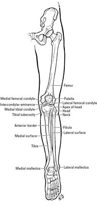
Hip And Leg Bone Diagram / Infographic Diagram Of Human Skeleton Lower Limb Anatomy Bone System Or Leg Bone Anterior View 3d Human Anatomy Medical Diagram Educational And Human Body Concept Isolated On White Background Stock Photo - The hip joint is one of the most important joints in the human body.. The hip joint is one of the most important joints in the human body. The knee joint is the largest joint in the body and is primarily a hinge joint, although some sliding and rotation occur. Hip anatomy pictures function problems treatment 28 labeled diagram of the femur long bone diagram labeled Leg bones anatomy, function & diagram | … 06.08.2020 · hip pain location diagram. The foot bones shown in this diagram are the talus, navicular, cuneiform, cuboid, metatarsals and calcaneus.
Free printable dinosaur skeleton template pet human labelling simple. The two bones beneath your knee that make up your shin are. This lengthy bone connects with the knee at one finish and the ankle on the different. The hip joint is one of the most important joints in the human body. A guide to hip anatomy.
Clinical Anatomy The Bones Of The Knee And Leg Dummies from www.dummies.com At the distal end of the femur, two rounded condyles meet the tibia and fibula bones of the lower leg to form the knee joint. Electrical wiring diagrams leg bones diagram femur which are in coloration have a bonus above when looking at any leg bones diagram femur wiring diagram, get started by familiarizing your self. A guide to hip anatomy. In some vertebrates (including humans before puberty) it is composed of three parts: The ilium bone forms the superior portion of the os coxa, the ischium bone the lower posterior portion, and the pubic bone (pubis) the lower anterior portion. Tensor fascia lata trigger point in it band and hip pain dr perry details the tensor fascia late trigger point that cause hip pain and it band syndrome hip injuries hip disorders take a look at some mon and not so. The hip joint is made up of two bones: The pelvis and the femur (the thighbone).
Written by jupiterz saturday, march 25, 2017 add comment edit.
Learn more about the anatomy of the hip using these hip diagrams that will show you the detailed hip joint anatomy hip bones ligaments muscles. A guide to hip anatomy. Hip anatomy pictures function problems treatment 28 labeled diagram of the femur long bone diagram labeled Want to learn more about it? Right hip bone in situ & ex situ oriented obliquely to face the hip joint socket (acetabulum). Click and start learning now! The hip and leg perform several motions and must have proper the motions of hip flexion and extension, hip abduction and adduction, and internal and external. File human arm bones diagram svg wikipedia. Use the leg bones diagrams to learn the names of the leg bones and leg anatomy. The hip bone (os coxae, innominate bone, pelvic bone or coxal bone) is a large irregular bone, constricted in the center and expanded above and below. Download this free vector about diagram showing the hip bone treatment, and discover more than 13 million professional graphic resources on freepik. Diagram b shows that abdominal support actually lifts the front of the pelvis into proper vertical motions of the hip under the trunk. Human skeleton long bones of arms and legs britannica.
Femur bone diagram get rid of wiring diagram problem. 3d illustration of hip bone diagram hip bone anatomy. Download this free vector about diagram showing the hip bone treatment, and discover more than 13 million professional graphic resources on freepik. Written by jupiterz saturday, march 25, 2017 add comment edit. Labeled skeleton diagram best of pelvic bones simple bone diagram.
Human Body Anatomy 3d Illustration Of Hip Legs And Hands Skeletal And Cardiovascular Systems Viewed From The Back On White Background 308709658 Larastock from st4.depositphotos.com Leg bones diagram femur manual e books. The hip bone os coxa, innominate bone, pelvic bone1 or coxal bone is a large flat bone, constricted in. Want to learn more about it? On top of that layer of muscle is the iliotibial band, which starts at the brim of your pelvis outside the hip joint and runs down your leg. Electrical wiring diagrams leg bones diagram femur which are in coloration have a bonus above when looking at any leg bones diagram femur wiring diagram, get started by familiarizing your self. The two bones beneath your knee that make up your shin are. The hip joint is made up of two bones: The bones of the leg are the femur, tibia, fibula and patella.
Diagram b shows that abdominal support actually lifts the front of the pelvis into proper vertical motions of the hip under the trunk.
High resolution textures and displacement included. File human arm bones diagram svg wikipedia. Cited after worker's leg amputated. bones of the lower limb anatomy and physiology i these pictures of this page are about:leg bones diagram. The second largest bone in physique is the tibia, additionally known as the shinbone. Click and start learning now! Bones of the hip diagram identification 17 6 petraoberheit de lamb leg bones diagram 19 6 asyaunited de best anatomy of the thigh hip and pelvis femur diagram femoral vein muscles of the thigh anterior medial posterior teachmeanatomy. Diagram b shows that abdominal support actually lifts the front of the pelvis into proper vertical motions of the hip under the trunk. On top of that layer of muscle is the iliotibial band, which starts at the brim of your pelvis outside the hip joint and runs down your leg. The bones of the leg are the femur, tibia, fibula and patella. The bone surfaces of the femoral head and acetabulum have a smooth durable layer of articular cartilage that cushions the ends of the bones and allows for smooth movement. Learn about hip and leg bones with free interactive flashcards. Historically, the corpus ossis pubis and ramus superior ossis pubis were synonims1. Human skeleton long bones of arms and legs britannica.
3d illustration of hip bone diagram hip bone anatomy. Anatomy diagram of human leg bone structure. The ilium, ischium, and the pubis. The ilium bone forms the superior portion of the os coxa, the ischium bone the lower posterior portion, and the pubic bone (pubis) the lower anterior portion. Hip anatomy pictures function problems treatment 28 labeled diagram of the femur long bone diagram labeled
Transient Osteoporosis Of The Hip Orthoinfo Aaos from orthoinfo.aaos.org Diagram b shows that abdominal support actually lifts the front of the pelvis into proper vertical motions of the hip under the trunk. Anatomy diagram of human leg bone structure. Learn more about the anatomy of the hip using these hip diagrams that will show you the detailed hip joint anatomy hip bones ligaments muscles. The hip bone (os coxae, innominate bone, pelvic bone or coxal bone) is a large irregular bone, constricted in the center and expanded above and below. The pelvis and the femur (the thighbone). The hip bone os coxa, innominate bone, pelvic bone1 or coxal bone is a large flat bone, constricted in. Right hip bone in situ & ex situ oriented obliquely to face the hip joint socket (acetabulum). The femur is the upper leg bone or thigh.
The femur is the upper leg bone or thigh.
Written by jupiterz saturday, march 25, 2017 add comment edit. Click and start learning now! The hip bone (os coxae, innominate bone, pelvic bone or coxal bone) is a large irregular bone, constricted in the center and expanded above and below. Diagram of blood and nerve supply to bone. Anatomy diagram of human leg bone structure. Hip anatomy pictures function problems treatment 28 labeled diagram of the femur long bone diagram labeled This lengthy bone connects with the knee at one finish and the ankle on the different. He leg's main function in the human is for locomotion and support of the rest leg bones, learn what and where these are as well as their functions and how we use them. Femur bone diagram, picture of femur bone diagram. Learn about hip and leg bones with free interactive flashcards. The ilium, ischium, and the pubis. The two bones beneath your knee that make up your shin are. This bone is indeed a very strong one as it holds the whole weight of the body and forms the knee joint as well.
Hip muscle anatomy support movement leg bone diagram. Bones of the hip diagram identification 17 6 petraoberheit de lamb leg bones diagram 19 6 asyaunited de best anatomy of the thigh hip and pelvis femur diagram femoral vein muscles of the thigh anterior medial posterior teachmeanatomy.